Describe 4 Methods Used to Study the Brain
The scientific method has six important steps. An Invasive imaging technique that provide color-coded images of brain activity by tracking the brains use of radioactively tagged compounds.

Into The Mind Of Flow Activate Your Subconscious Brain Images Brain Waves Flow State
Sometimes these two things are combined together.

. Commonly used brain imaging techniques are. Functional magnetic resonance imaging fMRI computerized tomography CT positron emission tomography PET electroencephalography EEG and. Modern technology that provides ways of studying the structure and function of the brain without the need of invasive techniques.
Here are some of the techniques used to study and help the human brain. For example multiple-voxel pattern analysis MVPA has been used to identify the subtle differences in activation patterns across voxels whereas neural connectivity analysis has been used to examine functional interactions among brain regions. Better known as an EEG this technique uses electrodes placed over the scalp to record the electrical activity of the brain with a particular focus on the cerebral cortex.
In the first fMRI study that used pattern analysis methods Haxby et al. Current research has used TMS to study the brain areas responsible for emotion and cognition and their roles in how people perceive intention and approach moral reasoning Kalbe et. While receiving the stimulations the patient is normally fully awake no anaesthetic.
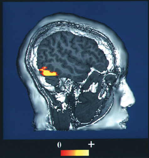
Procedure of PET Scan. New technologies experimental methods and animal experimentation have led to an increased understanding of. Your question should start with words like how what when where or why.
It is a relatively new discipline within medicine neuroscience and psychology. Functional imaging can be performed by two methods. Click card to see definition.
Some involve scanning the living brain looking for patterns of electrical activity associated. The fMRI creates images of brain structure and activity. The traditional method for studying cerebral lateralization is unilateral lesions study which is the study of the location and effect lesions.
These techniques ultimately have the same goal in that they aim to produce coherent representations of the brain. This method depicts the changes in brain. Recording Electrical Activity in the Brain.
A variety of methods are used by scientists in order to study the different areas and functions of the brain. Methods of Cognitive Neuroscience The cognitive neuroscience triangle can be used to categorize the methods of cognitive neuroscience used to study the relation between brain function and cognition. Computational Modeling Cognitive Neural 6.
The complex phenomenon of memory is explored by combining evidence from many areas of research. It is the test of choice to evaluate for the four types of. Persona lies in a scanner and performs a mental task.
This is more useful method to understand the behaviour from point of view of hereditary and environmental influences. These technological methods include the encephalogram EEG magnetic resonance imaging MRI functional magnetic resonance imaging fMRI and positron emission tomography PET. Cerebral Lateralization and Functionality There are several methods for studying cerebral lateralization.
Tap card to see definition. The brain is the main focus of neuroscience. Functional Imaging is a method which allows scientists to detect electrical and chemical activities of the brain.
The first method is known as Position Emission Tomography PET. Four methods in the forefront are the study of unilateral lesions sodium amytal dichotic listening and functional brain imaging. TMS is a magnetic method used to stimulate small regions of the brain.
Tap again to see term. FMRI allows visualization of brain areas with increased blood flow during a functional task or even during rest Figs. PET and CAT scans used to be the normal way for psychologists like Brian to look at their patients brains.
In some cases the result of the stroke is a specific lack of ability. Lesions allow the scientist to observe any loss of brain function that may occur. Studying the brain gives us important insights into the underlying foundations of our behaviour and mental processes.
During the procedure a magnetic field generator or coil is placed near the head of the person receiving the treatment. A Cross-sectional study in which the children of different age groups will be studied simultaneously b longitudinal study in which the same individual will be studied in different stages of life. Scans reveal intricate brain wiring reports on the.
The images are then developed on sensitive film. The fMRI is now the most commonly used method of learning about brain structure. Description While structural MRI and DTI both use MRI scanners to study anatomy fMRI uses the same technology to study physiology.
But they are not used as much. Scientists who study the brain using experiments are called neuroscientists and like all scientists they use the scientific method to answer their questions. In addition to lesion approaches it is also possible to learn about the brain by studying the electrical activity created by the firing of its neurons.
Click again to see term. For instance when an individual suffers a stroke a blood clot deprives part of the brain of oxygen killing the neurons in the area and rendering that area unable to process information. One approach primarily used with animals is to place detectors in the brain to study the responses of specific neurons.
Computed Tomography Scan CT Computed Tomography Scan CT scans use a series of X-ray beams passed through the head. Figure 415 fMRI Image. This method creates cross-sectional images of the brain and shows the structure of the brain but not its function.
2001 illustrated that. Physicians who specialize in the performance and interpretation of neuroimaging in the clinical setting. It can measure changes in synaptic activity in nerves which can help to diagnose.
The stimulation take approximately around 20 to 30 minutes. Glucose oxygen or a drug that has been injected into a persons bloodstream. This method is the most extensively used method of functional imaging.
Radioactively Tagged Compounds in PET scans. Neuroimaging or brain scanning includes the use of various techniques to directly or indirectly image the structure function or pharmacology of the brain. The study of memory incorporates research methodologies from neuropsychology human development and animal testing using a wide range of species.

Concept Mapping Concept Map Teaching Brain Based Learning

Right Brain Left Brain Poster Zazzle Com Left Brain Right Brain Brain Poster Brain Drawing
4 3 Psychologists Study The Brain Using Many Different Methods Introduction To Psychology 1st Canadian Edition

How The Brain Learns Training Industry
No comments for "Describe 4 Methods Used to Study the Brain"
Post a Comment